Skip to content
Diagnostic workstation · MRI

RadImages Workstation MRI

A diagnostic workstation for MRI with synchronised multi-series viewing, registration and fusion of sequences, DWI/ADC, perfusion, angiography, segmentation and structured reporting.

Optimised for neurological, musculoskeletal, oncological and cardiac MRI.

Documentation
RadImages Workstation MRI

Workstation MRI capabilities

From routine reading to quantitative analysis.

01

Multi-series comparison

Synchronised viewing of several sequences and planes.

02

Prior study comparison

Automatic alignment of current and previous studies.

03

Registration and fusion

Fuse T1, T2, FLAIR, DWI and PET with adjustable colour maps.

04

DWI and ADC

Diffusion analysis with ADC maps and ROI statistics.

05

Perfusion

Dynamic contrast and perfusion analysis with parametric maps.

06

MR angiography

MIP/MinIP, vessel tracking and stenosis assessment.

07

Segmentation and 3D

Manual and semi-automatic segmentation with volume rendering.

08

Measurements and ROI

Calibrated measurements, ROI statistics and annotations.

09

Structured reporting

Templates with automatic transfer of measurements into the report.

10

Cardiac MR package

Function, LGE and tissue characterisation.

11

Multi-monitor support

Layouts across several diagnostic monitors.

12

PACS integration

Direct connection to RadImages PACS and third-party archives.

13

Hanging protocols

Presets per protocol, body region and reader.

14

Cine playback

Dynamic series playback for cardiac and functional studies.

15

Data security

Personal accounts, permissions and an audit trail of every action.
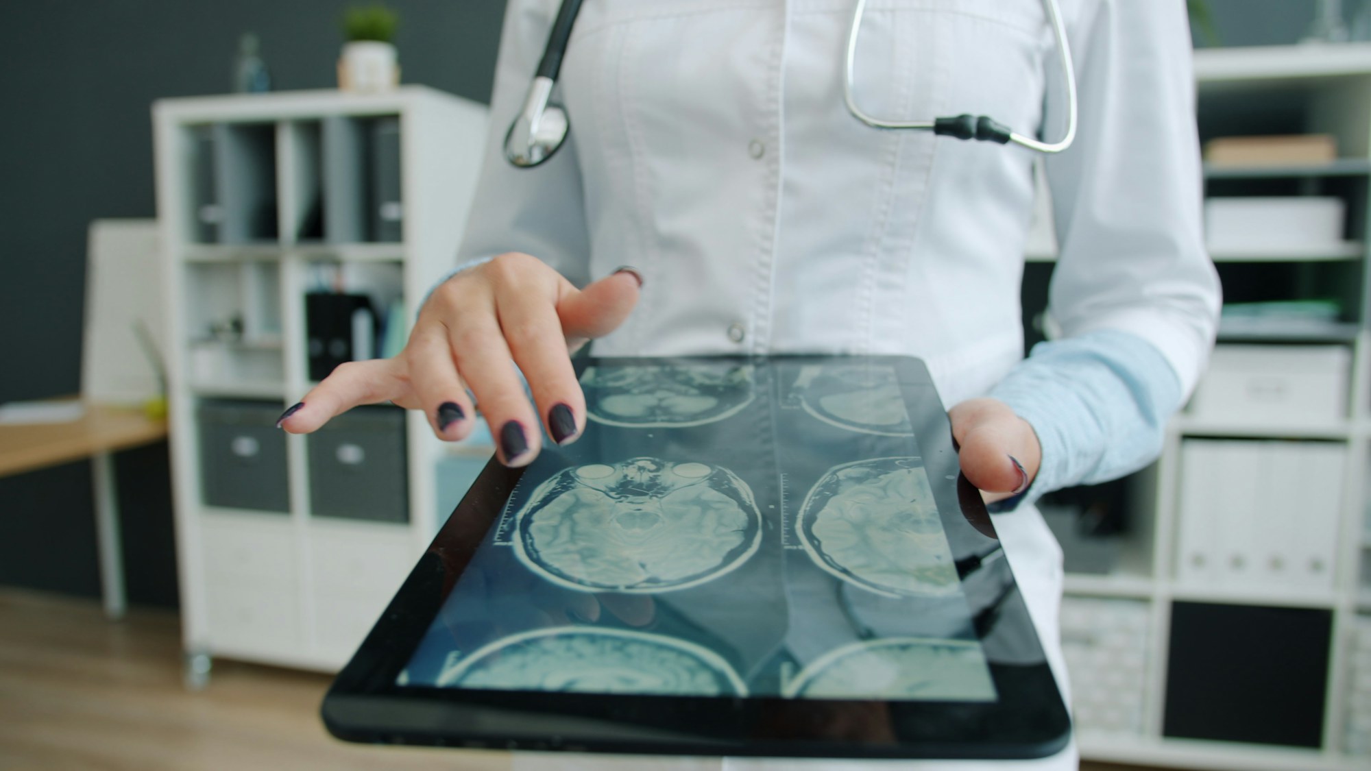
Who is Workstation MRI for?

MRI centres and radiology departments reading neuro, musculoskeletal, oncology and cardiac MRI.

  • MRI centres
  • Neuroradiology
  • Oncology
  • Cardiology
Clinical modules

Modules for Workstation MRI

Applications and options available with this solution. Select a group to see the details.

Pair 1 (T1 + T2 / Jet)

Fusion of T1 and T2 series in the axial plane with the Jet colormap.

Pair 1 (T1 + T2 / Jet)

Fusion of T1 and T2 series in the axial plane with the Jet colormap.

The image shows fusion of two MRI series in the axial plane: the base layer is T1 (AutoBind M3D/LAVA T1) in grayscale, and the overlay is T2 (AutoBind SSFSE T2) with the Jet colormap at 50% opacity. Jet maps T2 intensity from blue through green and yellow to red, enabling direct anatomical correlation of both sequences in one view.

Pilot project

Try RadImages free for 3 months

We deploy the system, connect your equipment and train your staff. You work in real conditions and decide with no obligations.

  • Deployment on-premise or in the cloud
  • Equipment connection and HIS/RIS integration
  • Training and support included
  • No payment, no obligations
3months free
About the pilot programme

MRI reading without compromise

Quantitative tools and fusion that used to require separate software, in one workstation.

RadImages products are software intended for use by medical professionals. All images, diagrams and illustrations on this website are provided for informational and illustrative purposes only and do not constitute medical advice.